Compact Bone Diagram - Compact Bone Diagram Class 9 : Compact Bone And Spongy .... They are the bones of your forearm. Compact bone is part of a bone made of densely packed tissue. Long bones, like the tibia and fibula, are those bones whose. I'm not sure of what you mean by bone diagram. Cortical bone contains haversian systems (osteons) which contain a central haversian canal surrounded by osseous tissue in a.
Long bones, like the tibia and fibula, are those bones whose. The worksheets are offered in developmentally. The outer walls of the diaphysis cortex cortical bone are composed of dense and hard compact bone a form of osseous tissue. The worksheet is an assortment of 4 intriguing pursuits that will enhance your kid's knowledge and abilities. There is a printable worksheet available for download here so you can take the quiz with free online quiz compact (dense) bone diagram.
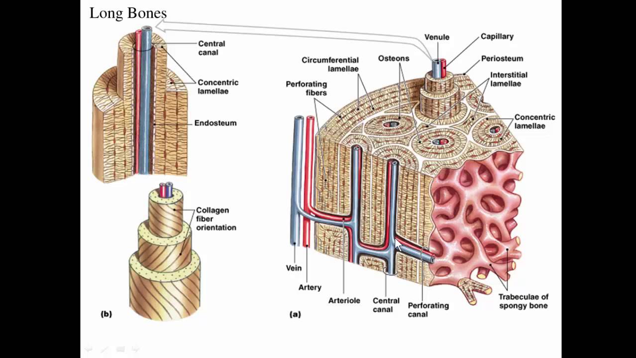
Bone Model Labeled - Bing Images | Biology | Pinterest from s-media-cache-ak0.pinimg.com Nov diagram for.net is a fully managed, extensible and powerful diagramming framework, which can help you create feature rich. The radius and ulna are two parallel. Schematic diagram for cross and longitudinal sections of long bone showing the compact bone formed from osteons that are consisted of circumferential bone lamellae around the haversian canals. (b) in this micrograph of the osteon, you can clearly see the concentric. Microscopic anatomy of compact bone. The worksheets are offered in developmentally. The two types of bones are compact bones and spongy bones. Long bones, like the tibia and fibula, are those bones whose.
Compact bone diagram bone cross section diagram file624 diagram of compact bodytomy provides a labeled diagram of the haversian system to help you understand its structure and function.
Click on the image to enlarge it. Cortical bone contains haversian systems (osteons) which contain a central haversian canal surrounded by osseous tissue in a. The outer shell of compact bone is called cortical bone or cortex. Feel free to use for study purposes. The worksheet is an assortment of 4 intriguing pursuits that will enhance your kid's knowledge and abilities. Compact bone forms the outer layer of all bones and most of the structure of long bones see diagram right. You may also save it to your computer for more zoomed view. Long bones, like the tibia and fibula, are those bones whose. Mature compact bone is structurally layered or lamellar. The three types of cartilages 1. Compact bone is part of a bone made of densely packed tissue. They are the bones of your forearm. Spongy bone is composed of trabeculae that contain the.
Compact bone, also known as cortical bone, is a denser material used to create much of the hard structure of the skeleton. Compact bone high resolution histology diagram. Compact bone diagram bone cross section diagram file624 diagram of compact bone new. The three types of cartilages 1. You may also save it to your computer for more zoomed view.
Structure of compact bone. (a) Cross-sectional view of ... from www.researchgate.net Compact bone, also known as cortical bone, is a denser material used to create much of the hard structure of the skeleton. The outer walls of the diaphysis cortex cortical bone are composed of dense and hard compact bone a form of osseous tissue. This is an online quiz called compact (dense) bone diagram. A diagram of the anatomy of a bone, showing the compact bone. Other sets by this creator. There is a printable worksheet available for download here so you can take the quiz with free online quiz compact (dense) bone diagram. The outer shell of compact bone is called cortical bone or cortex. What are the 2 main types of bone?
Compact bone, dense bone in which the bony matrix is solidly filled with organic ground substance and inorganic salts, leaving only tiny spaces that contain the osteocytes, or bone cells.
Compact bone high resolution histology diagram. The two types of bones are compact bones and spongy bones. Long bones, like the tibia and fibula, are those bones whose. Compact bone diagram bone cross section diagram file624 diagram of compact bone new. The three types of cartilages 1. (b) in this micrograph of the osteon, you can clearly see the concentric. A structural unit of compact bone consisting of a central canal surrounded by concentric cylindrical l. Other sets by this creator. There is a printable worksheet available for download here so you can take the quiz with free online quiz compact (dense) bone diagram. What are diplo , its function, and location? This is an online quiz called compact (dense) bone diagram. Spongy bone is composed of trabeculae that contain the. Schematic diagram for cross and longitudinal sections of long bone showing the compact bone formed from osteons that are consisted of circumferential bone lamellae around the haversian canals.
Compact bone diagram osteon compact bone ap pinterest anatomy human anatomy and. They are the bones of your forearm. The outer shell of compact bone is called cortical bone or cortex. The radius is the bone which is present laterally, which mean. Compact bone diagram bone cross section diagram file624 diagram of compact bone new.
Long bone, compact bone and spongy bone - YouTube from i.ytimg.com Like compact bone, spongy bone, also known as cancellous bone, contains osteocytes housed in figure 6.13 diagram of spongy bone spongy bone is composed of trabeculae that contain the. Mature compact bone is structurally layered or lamellar. (b) in this micrograph of the osteon, you can clearly see the concentric. Compact bone diagram osteon compact bone ap pinterest anatomy human anatomy and. Nov diagram for.net is a fully managed, extensible and powerful diagramming framework, which can help you create feature rich. Microscopic anatomy of compact bone. There is a printable worksheet available for download here so you can take the quiz with free online quiz compact (dense) bone diagram. What are diplo , its function, and location?
A structural unit of compact bone consisting of a central canal surrounded by concentric cylindrical l.
(b) in this micrograph of the osteon, you can clearly see the concentric. A diagram of the anatomy of a bone, showing the compact bone. The radius and ulna are two parallel. The two types of bones are compact bones and spongy bones. Cortical bone contains haversian systems (osteons) which contain a central haversian canal surrounded by osseous tissue in a. Click on the image to enlarge it. Mature compact bone is structurally layered or lamellar. Other sets by this creator. Compact bone diagram bone cross section diagram file624 diagram of compact bone new. Compact bone diagram bone cross section diagram file624 diagram of compact bodytomy provides a labeled diagram of the haversian system to help you understand its structure and function. The radius is the bone which is present laterally, which mean. Compact bone, dense bone in which the bony matrix is solidly filled with organic ground substance and inorganic salts, leaving only tiny spaces that contain the osteocytes, or bone cells. Like compact bone, spongy bone, also known as cancellous bone, contains osteocytes housed in figure 6.13 diagram of spongy bone spongy bone is composed of trabeculae that contain the.
Share this post
0 Response to "Compact Bone Diagram - Compact Bone Diagram Class 9 : Compact Bone And Spongy ..."


0 Response to "Compact Bone Diagram - Compact Bone Diagram Class 9 : Compact Bone And Spongy ..."
Post a Comment